About the Human Spine
Anatomy & Function
The spine is remarkable and complex, and a basic understanding of its anatomy and function is especially important for patients managing spinal disorders.
Spine Functions
The three main functions of the spine include:
- Protecting the spinal cord, nerve roots & several of the body’s internal organs
- Providing structural support & balance to maintain an upright posture
- Enabling flexible motion of the spine & body
Regions of the Spine
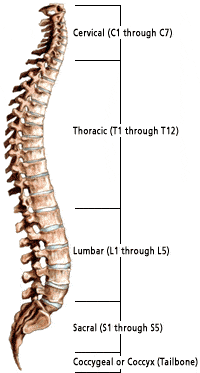
The spine is divided into four main regions: cervical, thoracic, lumbar and sacral. Each region has specific characteristics and functions.
Cervical Spine
The neck region of the spine is known as the cervical spine. It consists of seven vertebrae, abbreviated C1 through C7 (top to bottom). These vertebrae protect the brain stem and the spinal cord, support the skull and allow for a wide range of head movement.
The first cervical vertebra (C1) is called the atlas. The atlas is ring-shaped, and it supports the skull. C2 is called the axis. It is circular in shape with a blunt, peg-like structure (called the odontoid process or “dens”) that projects upward into the ring of the Atlas. Together, the atlas and axis enable the head to rotate and turn. The other cervical vertebrae (C3 through C7) are shaped like boxes with small spinous processes (finger-like projections) extending from the back of the vertebrae.
Thoracic Spine
Beneath the last cervical vertebra are the 12 vertebrae of the thoracic spine. These are abbreviated T1 through T12 (top to bottom). T1 is the smallest and T12 is the largest thoracic vertebra. The thoracic vertebrae are larger than the cervical bones and have longer spinous processes.
In addition to longer spinous processes, rib attachments add to the thoracic spine’s strength. These structures make the thoracic spine more stable than the cervical or lumbar regions. The rib cage and ligament systems also limit the thoracic spine’s range of motion and protect many vital organs.
Lumbar Spine
The lumbar spine has five vertebrae abbreviated L1 through L5 (largest). The size and shape of each lumbar vertebra is designed to carry most of the body’s weight. Each structural element of a lumbar vertebra is bigger, wider and broader than similar components in the cervical and thoracic regions.
The lumbar spine has more range of motion than the thoracic spine but less than the cervical spine. The lumbar facet joints allow for significant flexion and extension movement while limiting rotation.
Sacral Spine
The sacrum is located behind the pelvis. Five bones (abbreviated S1 through S5), fused into a triangular shape, form the sacrum. The sacrum fits between the two hipbones connecting the spine to the pelvis. The last lumbar vertebra (L5) articulates (moves) with the sacrum. Immediately below the sacrum are five additional bones fused together to form the coccyx (tailbone).
Pelvis & Skull
Although not typically viewed as part of the spine, the pelvis and the skull are anatomic structures that closely interrelate with the spine and have a significant impact on a person’s balance.
Spinal Planes
To help further understand and describe the anatomy, spine specialists often refer to specific body planes. A body plane is an imaginary flat, two-dimensional surface that is used to define a particular anatomical area.
Table 1
| Term | Meaning |
| Frontal or Coronal Plane | Divides the front and back halves of the entire body. |
| Median or Sagittal Plane | Divides the left and right sides of the entire body. |
| Transverse or Axial Plane | Divides the body at the waist (top and bottom halves of the body). |
Spinal Curves
When viewed from the front (coronal plane), the healthy spine is straight. A sideways curve in the spine is known as scoliosis. When viewed from the side (sagittal plane) the mature spine has four distinct curves. These curves are described as being either kyphotic or lordotic.
A kyphotic curve is a convex curve in the spine (i.e. convexity toward the back of the spine). The curves in the thoracic and sacral spine are kyphotic.
A lordotic curve is concave (i.e. concavity towards the back of the spine) and is found in the cervical and lumbar levels of the spine.
Vertebral Structures
All vertebrae consist of the same basic elements, with the exception of the first two cervical vertebrae. The outer shell of a vertebra is made of cortical bone. This type of bone is dense, solid and strong. Inside each vertebra is cancellous bone, which is weaker than cortical bone and consists of loosely knit structures that look somewhat like a honeycomb. Bone marrow, which forms red blood cells and some types of white blood cells, is found within the cavities of cancellous bone.
Vertebrae consist of the following common elements:
- Vertebral body: The largest part of a vertebra. When viewed from above, it has a somewhat oval shape. When viewed from the side, the vertebral body is shaped like an hourglass: thicker at the ends and thinner in the middle. The body is covered with strong cortical bone and filled with cancellous bone.
- Pedicles: Two short processes made of strong cortical bone and protruding from the back of the vertebral body.
- Laminae: Two relatively flat plates of bone extending from the pedicles on either side and joining in the midline.
- Processes: There are three types of processes: articular, transverse and spinous. The processes serve as connection points for ligaments and tendons. The four articular processes link with the articular processes of adjacent vertebrae to form the facet joints. The facet joints, combined with intervertebral discs, allow for motion in the spine. The spinous process extends posteriorly from the point where the two laminae join, acting as a lever to effect motion of the vertebra. The transverse processes are the small, bony pieces protruding from the right and left side of each vertebra. These processes serve as an attachment for muscle and ligaments and act as a point of articulation of the thoracic ribs.
- Endplates: The top (superior) and bottom (inferior) of each vertebral body is “coated” with an endplate. Endplates are complex structures that blend into the intervertebral disc and help support the disc.
- Intervertebral foramen: The pedicles have a small notch on their upper surface and a deep notch on their bottom surface. When the vertebrae are stacked on top of one another, the pedicle notches form an area called the intervertebral foramen. This area is of critical importance, since the nerve roots exit from the spinal cord through this area to the rest of the body.
Facet Joints
The joints in the spinal column are located posterior to the vertebral body (on the back side). These joints help the spine bend, twist and extend in different directions. Although the joints enable movement, they also restrict excessive movements like hyperextension and hyperflexion (i.e. whiplash).
Each vertebra has two facet joints. The superior articular facet faces upward and works like a hinge with the inferior articular facet (below).
Like other joints in the body, each facet joint is surrounded by a capsule of connective tissue and produces synovial fluid to nourish and lubricate the joint. The surfaces of the joint are coated with cartilage to help each joint move (articulate) smoothly.
Intervertebral Discs
Between the vertebral bodies is a "cushion" called an intervertebral disc. Each disc absorbs the stress and shock the body incurs during movement and prevents the vertebrae from grinding against one another. The intervertebral discs are the largest structures in the body without a vascular supply. Through osmosis, each disc absorbs needed nutrients.
Each disc is made up of two parts: the annulus fibrosis and the nucleus pulposus.
Annulus Fibrosus
The annulus is a sturdy, tire-like structure that encases a gel-like center—the nucleus pulposus. The annulus enhances the spine’s rotational stability and helps resist compressive stress.
The annulus consists of water and layers of sturdy elastic collagen fibers. The fibers are oriented at different horizontal angles, similar to the construction of a radial tire. Collagen gains its strength from strong fibrous bundles of protein that are linked together.
Nucleus Pulposus
The center portion of each intervertebral disc is a filled with a gel-like elastic substance. Together with the annulus fibrosus, the nucleus pulposus transmits stress and weight from vertebra to vertebra. Like the annulus fibrosus, the nucleus pulposus consists of water, collagen and proteoglycans. However, the proportion of these substances in the nucleus pulposus is different. The nucleus contains more water than the annulus.
The Spinal Cord and Nerve Roots
The spinal cord is a slender, cylindrical structure about the width of the little finger. The spinal cord begins immediately below the brain stem and extends to the first lumbar vertebra (L1). Thereafter, the cord blends with the conus medullaris that becomes the cauda equina, a group of nerves resembling the tail of a horse. The spinal nerve roots are responsible for stimulating movement and feeling. The nerve roots exit the spinal canal through the intervertebral foramen, which are small openings between each vertebra.
The brain and the spinal cord make up the central nervous system (CNS). The nerve roots that exit the spinal cord/spinal canal branch out into the body to form the peripheral nervous system (PNS).
Between the front and back portions of the vertebra (i.e. the mid-region) is the spinal canal housing the spinal cord and the intervertebral foramen. The foramen are small openings formed between each vertebra. These “holes” provide space for the nerve roots to exit the spinal canal and further branch out to form the peripheral nervous system.
Table 2
| Type of Neural Structure | Role/Function |
| Brain Stem | Connects the spinal cord to other parts of the brain. |
| Spinal Cord | Carries nerve impulses between the brain and spinal nerves. |
| Cervical Nerves (8 pairs) | These nerves supply the head, neck, shoulders, arms, and hands. |
| Thoracic Nerves (12 pairs) | Connects portions of the upper abdomen and muscles in the back and chest areas. |
| Lumbar Nerves (5 pairs) | Feeds the lower back and legs. |
| Sacral Nerves (5 pairs) | Supplies the buttocks, legs, feet, anal and genital areas of the body. |
| Dermatomes | Areas on the skin surface supplied by nerve fibers from one spinal root. |
Table 3
| Ligament Name | Description |
| Anterior Longitudinal Ligament (ALL) A primary spine stabilizer |
About 1 inch wide, the ALL runs the entire length of the spine from the base of the skull to the sacrum. It connects the front (anterior) of the vertebral body to the front of the annulus fibrosis. |
| Posterior Longitudinal Ligament (PLL) A primary spine stabilizer |
About 1 inch wide, the PLL runs the entire length of the spine from the base of the skull to sacrum. It connects the back (posterior) of the vertebral body to the back of the annulus fibrosis. |
| Supraspinous Ligament | This ligament attaches the tip of each spinous process to the other. |
| Interspinous Ligament | This thin ligament attaches to another ligament, called the ligamentum flavum, that runs deep into the spinal column. |
| Ligamentum Flavum The strongest ligament |
This yellow ligament is the strongest one. It runs from the base of the skull to the pelvis, in front of and behind the lamina, protecting the spinal cord and nerves. The ligamentum flavum also surrounds the facet joint capsules. |
Muscles and Tendons
The muscular system of the spine is complex, with several different muscles playing important roles. The primary function of the muscles is to support and stabilize the spine. Specific muscles are associated with movement of specific parts of the anatomy. For example, the sternocleidomastoid muscle assists with movement of the head, while the psoas major muscle is associated with flexion of the thigh.
Muscles, either individually or in groups, are supported by fascia. Fascia is strong connective tissue. The tendon that attaches muscle to bone is part of the fascia. The muscles in the vertebral column are called flexors, rotators, or extensors.
Learn More About the Spine
To learn more about the anatomy and function of the brain, take advantage of these reputable resources:
Princeton Brain, Spine & Orthopedics knows that many of our patients like to research their condition and treatment
and look for useful resources on the internet. However, many find this a frustrating experience.
Princeton Brain, Spine & Orthopedics is pleased to offer you a unique health and wellness search service for internet
information that:
- Allows you to be an intelligent efficient user of the internet for health information
- Allows you to
find resources for top health and wellness sites in one place that are relevant to your
questions - Allows you to find resources in formats that you can "digest" and use to learn
What do you want to learn about?
To schedule an appointment for spine symptoms or spine disorders, call a Princeton Brian & Spine staff member at 609.921.9001 in New Jersey or 215.741.3141 in Pennsylvania. For your convenience, you can also request an appointment staff online.
Request an Appointment
Submit an appointment request on our patient portal or contact our New Jersey and Pennsylvania campuses to speak with a patient advocate.
Schedule an Appointment